omohyoid muscle ultrasound
We present a case of omohyoid muscle syndrome diagnosed based on the clinical presentation and a dynamic imaging study. The omohyoid muscle is a key to finding the suprascapular nerve when it is hard to find.

Pin Page
The geniohyoid muscle is one of the suprahyoid muscles of the neck that is innervated by the ventral ramus of C1.

. While we did not appreciate any variations in the OM there are several reported variations of the omohyoid muscle Rai et al 2008. Omohyoid muscle syndrome is a rare cause of an X-shaped bulging lateral neck mass that occurs on swallowing. The aim of this study was to obtain normative ultrasonography US data on the suprascapular nerve SSN and omohyoid muscle OM in the lateral cervical region.
This is a diagnostic case report of a 22-year-old mixed martial arts athlete who acquired this condition. Complications The major complication in the treatment of patients thought to have omohyoid syndrome is wrong diagnosis. Geniohyoid draws the hyoid bone up and forward during mastication and assists the opening of the mandible.
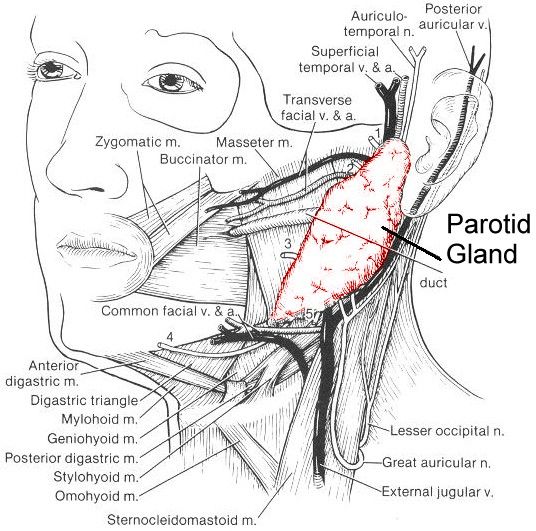
The placement of a central venous access into the internal jugular veinIJV is a commonly practiced clinical technique that allows fluid and blood transfusion and other rescue treatments. Surgical transection can achieve cure but due to limited studies available they should be reserved for patients who are extremely bothered. They are all attached to the hyoid bone and look like a strap.
It is a group of four pairs of muscles in the anterior part of the neck. We describe a chronic cerebro-spinal venous insufficiency patient who presented a omohyoid muscle entrapment of the internal jugular vein confirmed by both magnetic resonance venography and ultrasound investigation. Treatment is conservative and includes non-steroidal anti-inflammatories and muscle relaxants.
Botulinum toxin was injected into the inferior belly of one omohyoid muscle and the neck mass resolved. Omohyoid muscle syndrome has a characteristic feature of a protruding lateral neck mass during swallowing. Physical therapy including heat ice ultrasound and.
Real-time ultrasonography establishes the diagnosis demonstrating the anterolateral displacement of the sternocleidomastoid muscle by a thickened omohyoid muscle during swallowing. The SSN and OM are known to be related throughout the nerves course yet little imaging data exists on these structures at areas more proximal than the suprascapular foramen. The purpose of this study is to explore the anatomical relationship between the omohyoid muscle and the internal jugular vein on ultrasound guidance and.
Mylohyoid lineridge on the internal surface of the mandible. The other strap-muscles are not drawn in this illustration. The infrahyoid neck is separated from the suprahyoid neck by the hyoid bone arrow.
However the omohyoid muscle which is adjacent to the internal jugular vein is a rarely mentioned muscle of the infrahyoid muscles group. Also the cleidoatlanticus muscle which is variably present and courses from the clavicle vertically through the lateral cervical region to attach to the transverse process of the atlas has also been reported to alter the ultrasound. A omohyoid muscle surgical transection together with a patch angioplasty was performed.
Studies on internal jugular vein catheterization. The Omohyoid Syndrome is usually caused by trauma such as whiplash or by excessive vomiting. Authors Yun Yang 1 Xinqiang Wang 1 Weiliang Mao 1 Tongyun He 1 Zhaodong Xiong 2 Affiliations.
MRI scans of the neck can look for injury to the muscle and to exclude other causes such as tumors. Inferior mental spine of the mandible also known as the genial tubercle. The purpose of this study is to explore the anatomical relationship between the omohyoid muscle and the internal jugular vein on ultrasound guidance and provide a theoretical reference for jugular.
It is widely used to treat critically ill patients with. This technique can also be utilized for injection of botulinum toxin into the omohyoid muscle. Anatomical relationship between the omohyoid muscle and the internal jugular vein on ultrasound guidance BMC Anesthesiol.
They run closely together from the lateral neck toward the scapular notch. Anterior and middle fibers insert into a midline raphe that extends from the mandibular symphysis to the body of the hyoid bone the posterior fibers extend directly to. The sternothyroid sternohyoid thyrohyoid and omohyoid muscles.
An elongated transverse process can be defined as one that extends more laterally than the normal cervical transverse process lateral mass. 2012 Wiley Periodicals Inc. Ultrasound guidance may improve the accuracy of needle placement and decrease the incidence of needle-related complications.
2022 Jun 1322 1181.

Pin Page

Pin De Rubi Moreno En Rx Ultrasonido Radiologia Imagenologia
Pin Page

Muscles Anatomy Physiology Health Fitness Training Muscle Veins Medical Anatomy Anatomy Arteries Anatomy

Pin Page

This School Is Closed Right Now

Pin Page

Pectoral Clavipectoral And Axillary Fasciae Anatomy Thoracoacromial Artery Coracoid Process C Human Heart Anatomy Upper Limb Anatomy Subscapularis Muscle

Pin Page

Pin Page
Pin Page

Pin Page

Pin Page

Organization Of The Neck Flashcards Memorang

Reasonably Well

Pin Page

Pin Page

A Simple Interscalene Block The Manani S Technique Some Elements Of Distinction From Supraclavicular Perivascular Techniques

Tim Luijkx Timluijkx Human Body Anatomy Female Reproductive System Anatomy Skull Anatomy


Comments
Post a Comment